Molecular diagnostics examines DNA, RNA, and related biological signals to help identify infections, inherited conditions, and changes associated with disease. The answer to “what technologies are used in molecular diagnostics” depends on the target, sample, and clinical question. A nasal swab, for example, may be tested for a pathogen’s genetic material, while a blood sample may be examined for specific mutations. The instruments and workflows can look very different.
Polymerase chain reaction (PCR) and real-time PCR amplify selected genetic sequences, making small amounts easier to detect. Digital PCR partitions a sample into many reactions for more precise target measurement. Sequencing technologies can read genetic material across targeted regions or much broader portions of a genome. Other approaches include isothermal amplification, nucleic acid hybridization, and microarrays. Each has trade-offs in speed, sensitivity, cost, and the information it provides. No method fits every case.
The technology alone does not determine a reliable result. Sample quality, contamination controls, assay design, and laboratory validation matter too. A positive signal may need interpretation alongside symptoms, medical history, and other test findings. That nuance is easy to overlook. This guide introduces the main technology families, explains how their workflows differ, and outlines what to consider when comparing them. It is not a substitute for clinical advice, and test performance can vary by assay and setting. Even a sophisticated platform has limits. Understanding those limits is part of using molecular results responsibly.
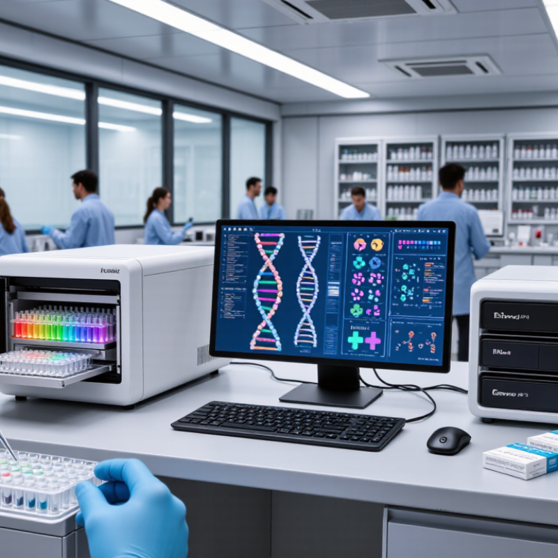
Molecular diagnostics reads DNA or RNA to detect pathogens, inherited variants, or changes linked to disease. The core principle is target recognition: primers or probes bind chosen genetic sequences. PCR then copies a target, while real-time PCR tracks amplification through fluorescence. Isothermal methods amplify nucleic acid at a steady temperature. Sequencing can examine many positions at once. Each approach depends on clean extraction, specific binding, and reliable controls.
The National Human Genome Research Institute’s DNA Sequencing Costs database shows sequencing costs fell from about $100 million per human genome in 2001 to below $1,000 by 2015. This reflects a major technology shift, not the price of every diagnostic test. In practice, sample quality, target abundance, and contamination can shape results. A negative result may reflect timing or a poor specimen, not just absence of disease. Not every sample behaves neatly.
Tips: Check that the specimen type and collection time match the test instructions. Look for internal controls, which help reveal extraction or amplification failure. And remember: a precise result still needs clinical context.
| Technology | Core principle | Typical targets and uses | Key strengths | Important considerations |
|---|---|---|---|---|
| Polymerase chain reaction (PCR) | Uses repeated temperature cycles and sequence-specific primers to amplify a selected DNA region. | Detection of specific DNA sequences, including pathogen genes and inherited variants. | Highly targeted and sensitive; established workflows and broad clinical use. | Requires thermal cycling and careful contamination control. Standard PCR generally indicates whether a target was amplified, not its starting quantity. |
| Real-time PCR (qPCR) | Measures fluorescence during amplification to monitor the accumulation of PCR products. | Quantification or detection of DNA targets; commonly used with reverse transcription to test RNA targets. | Combines amplification and signal measurement in a closed reaction vessel; supports quantitative analysis. | Results depend on assay design, controls, and calibration. Inhibition or poor sample quality can affect measurements. |
| Reverse-transcription PCR (RT-PCR) | Converts RNA into complementary DNA (cDNA), which can then be amplified by PCR. | RNA viruses, gene expression studies, and other RNA-based targets. | Enables PCR-based testing of RNA, which cannot be directly amplified by standard DNA polymerases. | RNA is vulnerable to degradation; extraction quality and reverse-transcription efficiency can influence results. |
| Digital PCR (dPCR) | Partitions a sample into many small reactions, classifies each as positive or negative, and estimates target concentration from the fraction of positive partitions. | Low-abundance DNA or RNA targets, rare variants, and precise nucleic-acid quantification. | Can provide absolute quantification without a standard curve and can detect low-frequency targets in suitable assays. | Partitioning systems and analysis add complexity; performance depends on assay design, sample quality, and adequate partition counts. |
| DNA sequencing | Determines the order of nucleotide bases. Methods include targeted sequencing and massively parallel sequencing. | Inherited variants, tumor-associated alterations, pathogen identification, and analysis of multiple genes or genomic regions. | Can identify sequence changes across one or many targets; broad sequencing can reveal variants not specified in advance. | Requires interpretation and quality review. Coverage, turnaround time, cost, and detection limits vary by method and assay. |
| Nucleic-acid hybridization and microarrays | Detects target sequences through binding, or hybridization, to complementary probes; arrays use many probes in parallel. | Known genetic variants, gene-expression patterns, and selected pathogen or genomic targets. | Can test many predefined sequences simultaneously, especially in array-based formats. | Usually best suited to targets represented by the probes; sequence changes can reduce binding and affect detection. |
| Fluorescence in situ hybridization (FISH) | Fluorescently labeled DNA probes bind to complementary sequences within fixed cells or tissue, allowing signals to be viewed microscopically. | Chromosome number or structure changes, gene rearrangements, and gene amplification in cells or tissue. | Shows where a genetic target is located and can preserve cellular context. | Typically evaluates selected targets rather than sequencing an entire genome; interpretation requires suitable specimens and microscopy expertise. |
| Isothermal nucleic-acid amplification | Amplifies nucleic acids at a constant temperature using specialized enzymes and primer designs, rather than repeated thermal cycling. | Rapid testing for selected DNA or RNA targets, including some infectious agents. | Can simplify equipment needs and support faster workflows for appropriately designed assays. | Primer design and nonspecific amplification require careful control; performance and validation are assay-specific. |
| CRISPR-based detection | Sequence-guided CRISPR-associated enzymes recognize a target; some systems generate a detectable signal after target recognition, often alongside a separate amplification step. | Research and developing diagnostic assays for selected pathogen or genetic targets. | Programmable sequence recognition can provide a flexible detection approach. | Clinical use, sensitivity, workflow, and regulatory status vary by assay; many approaches still require sample preparation or target amplification. |
It examines DNA or RNA to find pathogens, inherited variants, or changes linked to disease. Results depend on the sample.
PCR copies a selected DNA region many times. For RNA targets, a conversion step makes DNA first. Small signals matter.
It tracks DNA amplification using fluorescence. A bright signal is not proof by itself; controls and interpretation still matter.
A probe binds a matching genetic sequence. Fluorescent markers can make that binding measurable. Even one genetic change may affect the match.
Sequencing reads DNA bases across selected regions or broader areas. It may find changes a specific probe was not designed to detect.
Poor collection, contamination, inhibitors, or low target levels can affect results. Not every sample behaves neatly.
The sample may have been collected too early, or it may contain too little target material. A negative result does not always settle the question.
Confirm the specimen type and collection time match instructions. Internal, positive, and negative controls can reveal problems. No method is perfect.
What technologies are used in molecular diagnostics? These methods examine genetic material, such as DNA and RNA, to detect biological markers and help identify disease-related changes. The process begins with preparing a biological sample: cells are collected, nucleic acids are extracted, and unwanted material is removed so the genetic targets can be analyzed reliably.
When a target is present in small amounts, amplification methods make many copies to improve detection. Probes can then bind to specific sequences, while sequencing can read genetic material in greater detail. Different platforms interpret these signals in different ways, including measuring fluorescence, detecting changes in electrical signals, or analyzing sequence data. Together, sample preparation, amplification, target recognition, and result interpretation form a flexible toolkit for molecular analysis. The choice of technology depends on the question being asked, the sample type, and the level of detail required.

 BioX Medical