Advanced Professional Surgical Imaging System for Precise Diagnostics

Customization: Available
Classification: Physiological Functions of Diagnosis and Monitoring Equipment
Type: Surgical Fluorescence Imaging System

Product Description

📋

Basic Information

Model NO.:FLl-10B
Certification:CE, FDA, ISO13485
Monitor Size:27 Inches / 32 Inches
Resolution:1920*1080P
HS Code:9018903010
🔬

Product Description

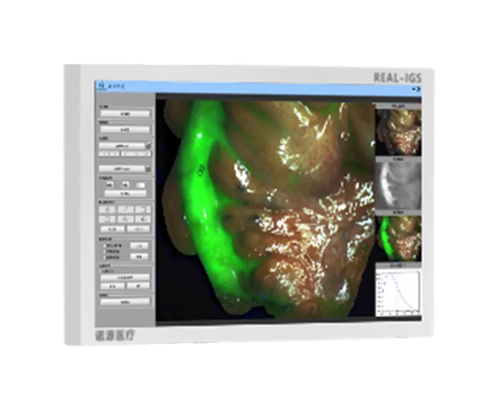
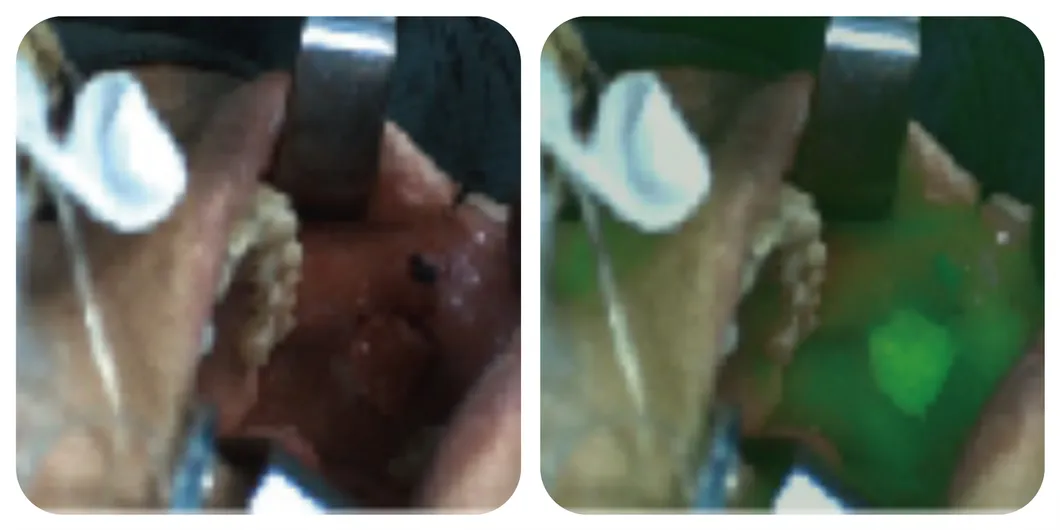
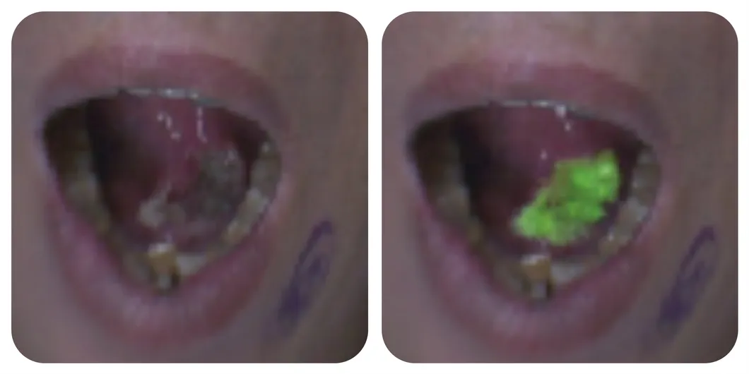
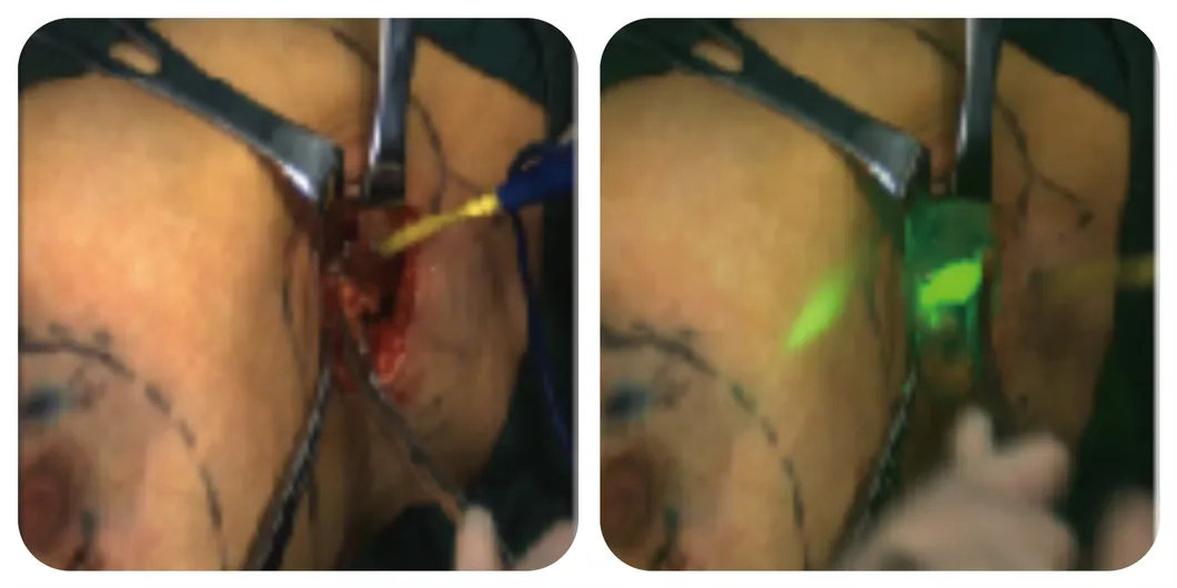
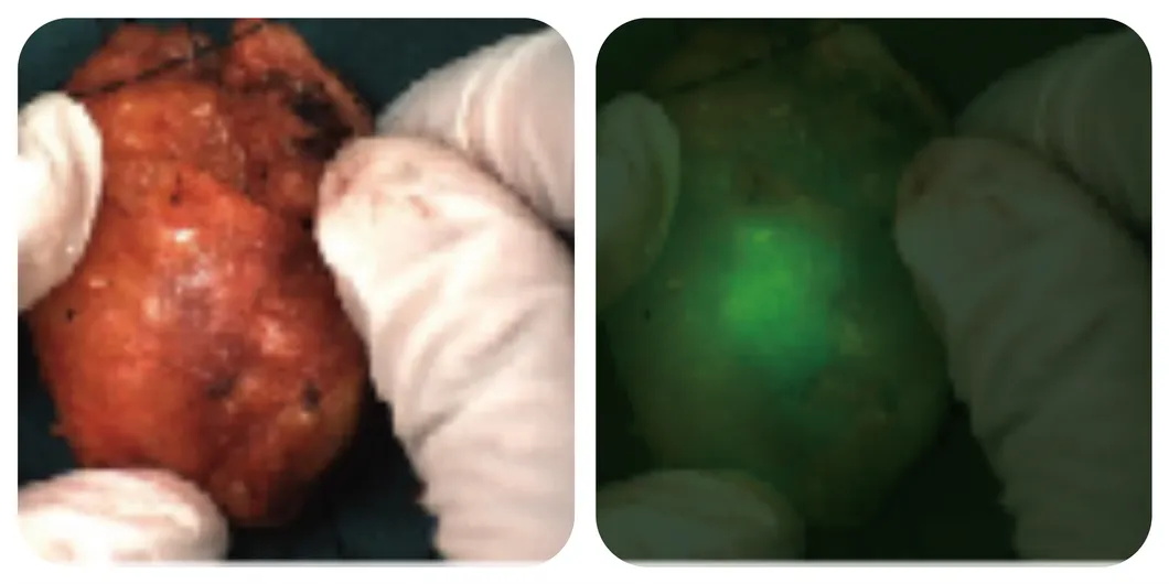
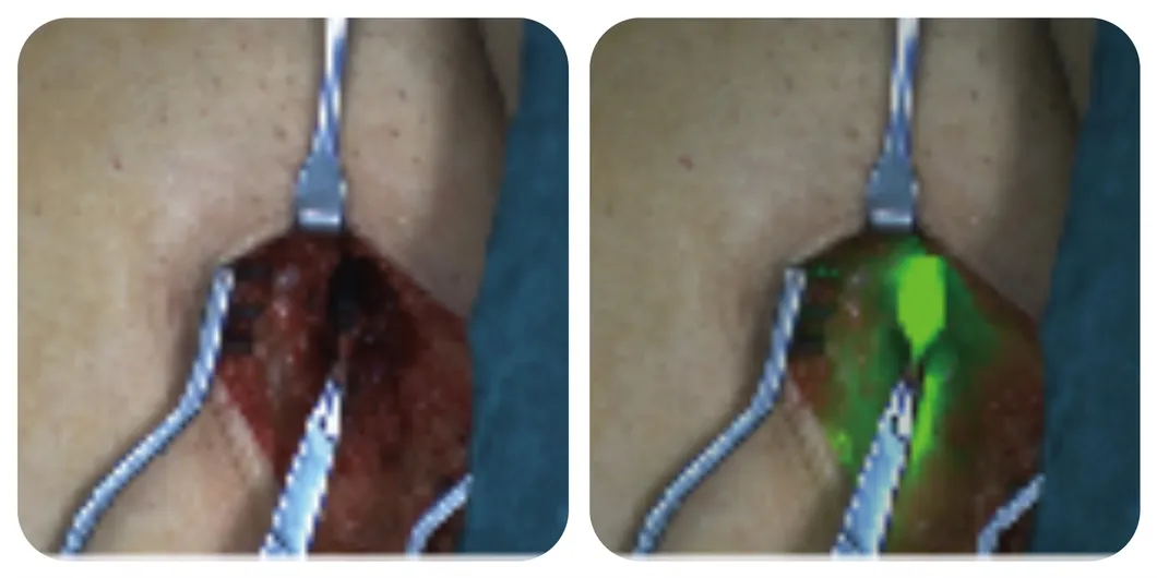
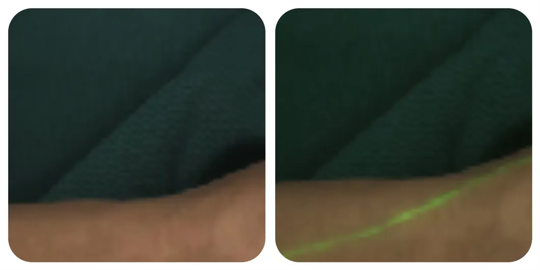
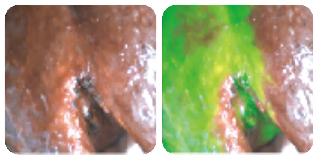
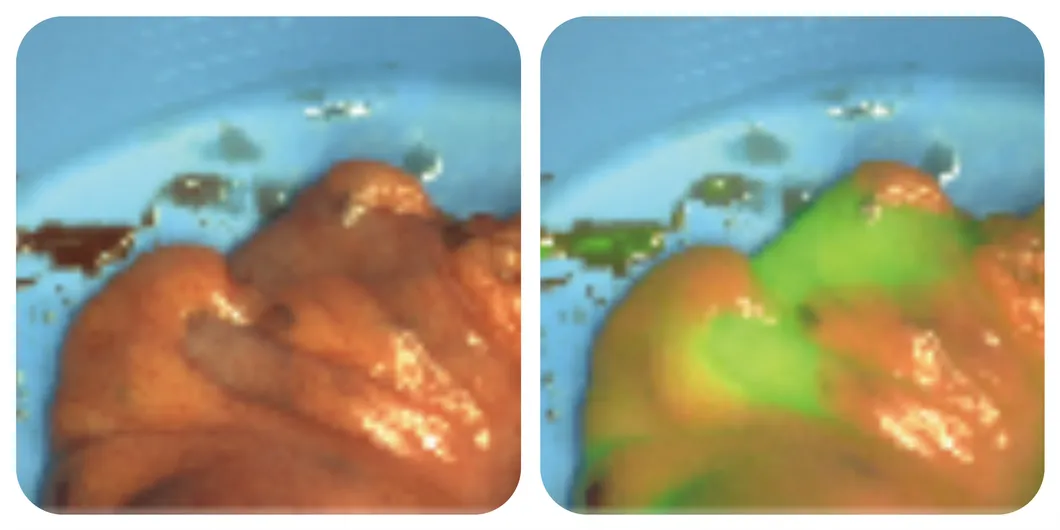
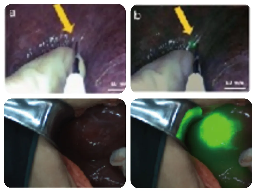
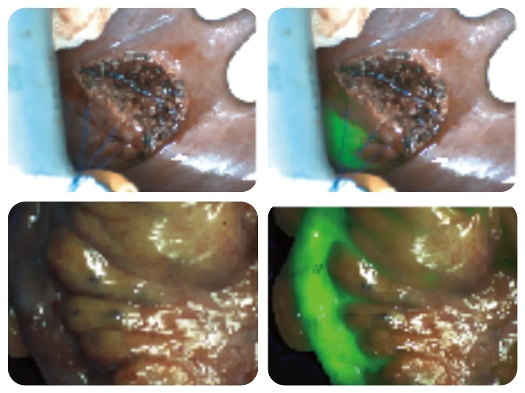
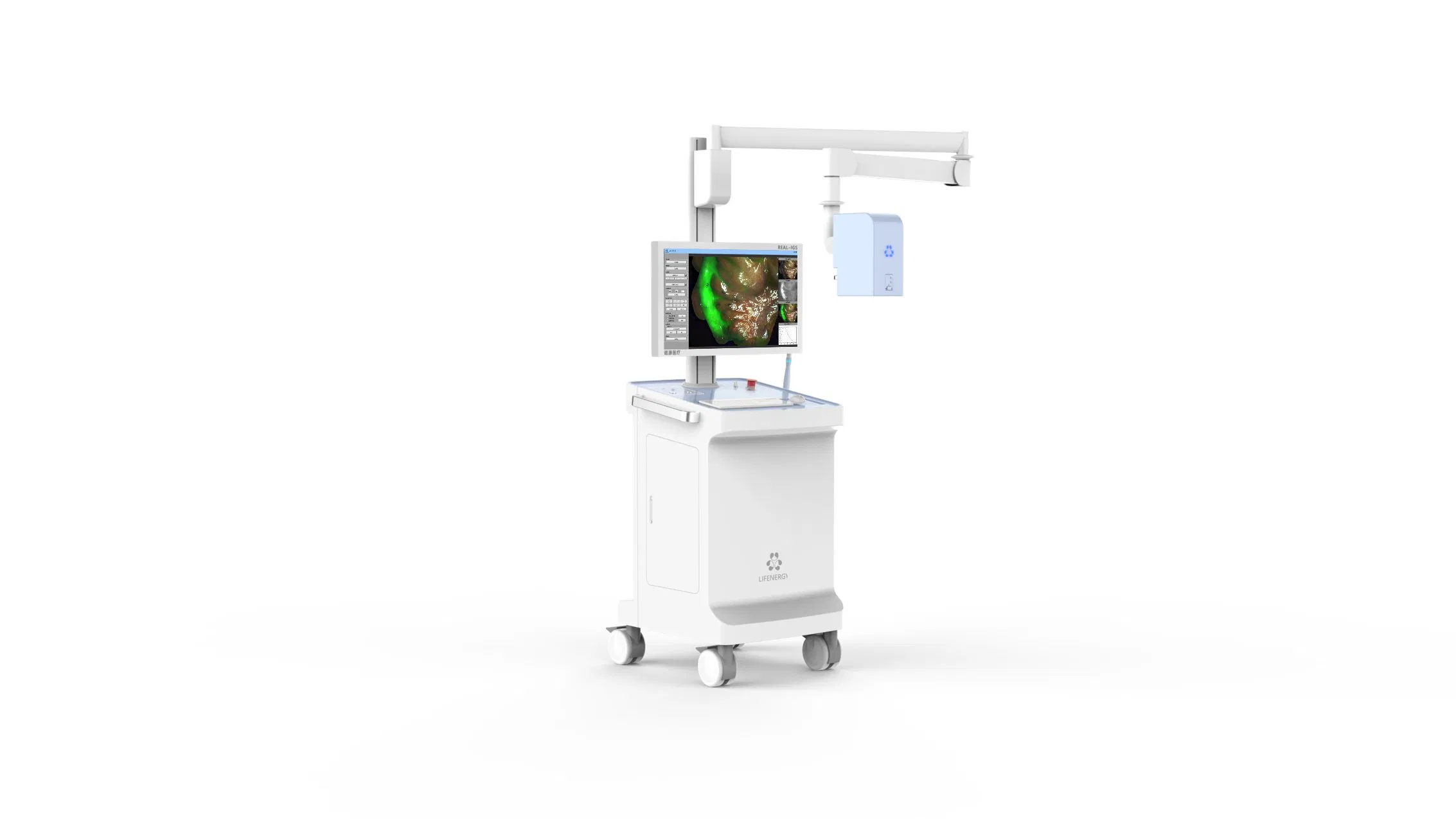
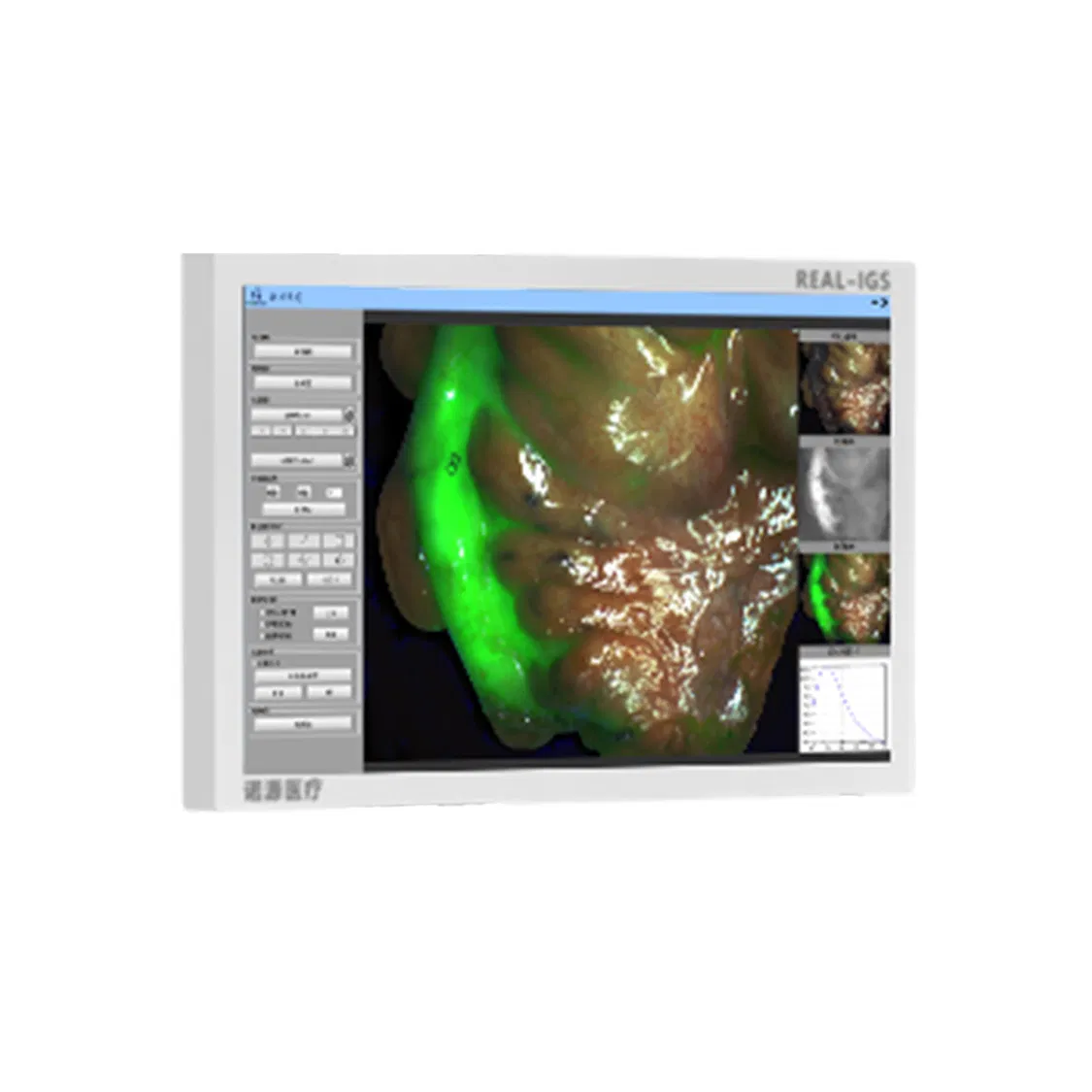
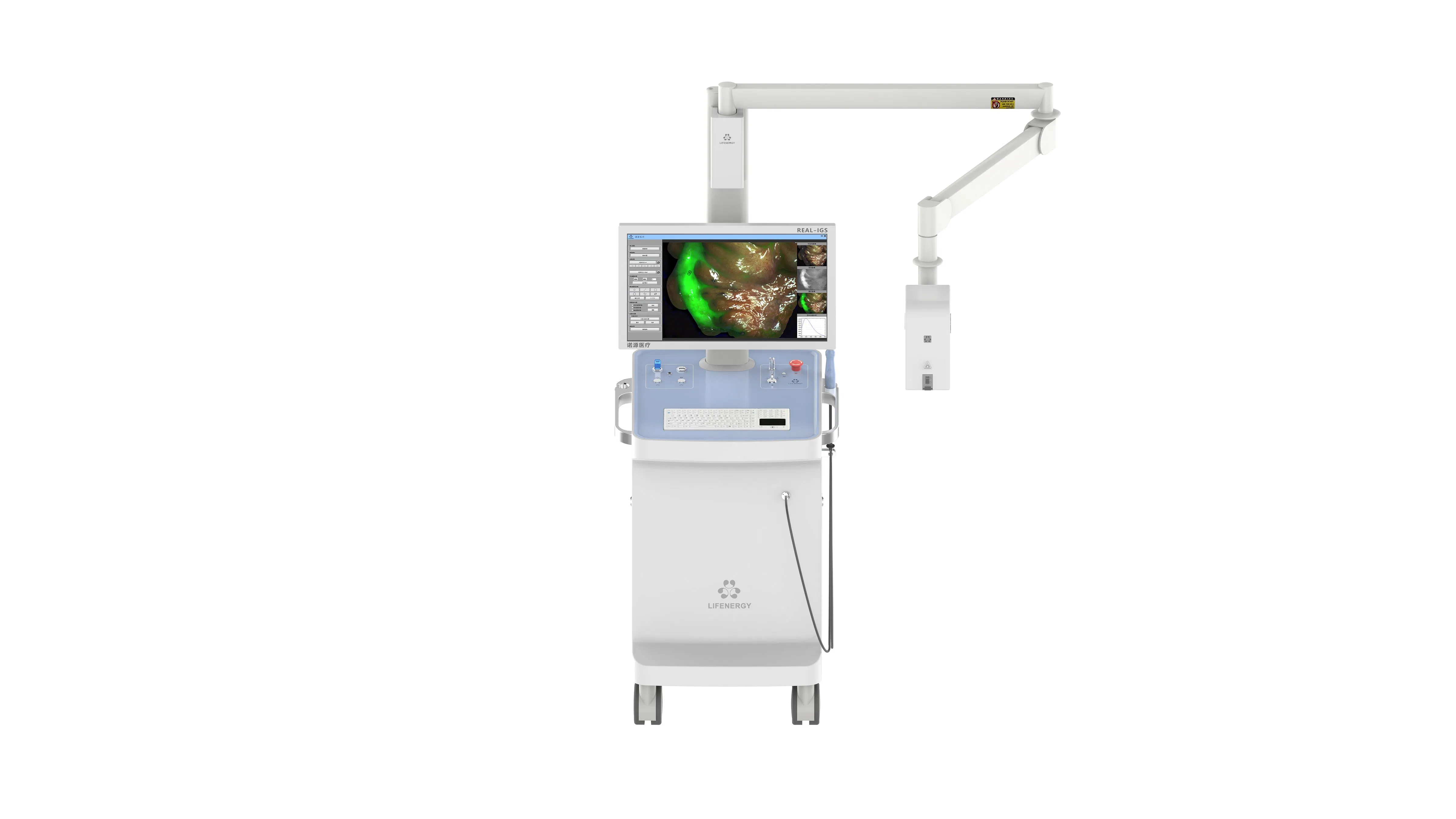
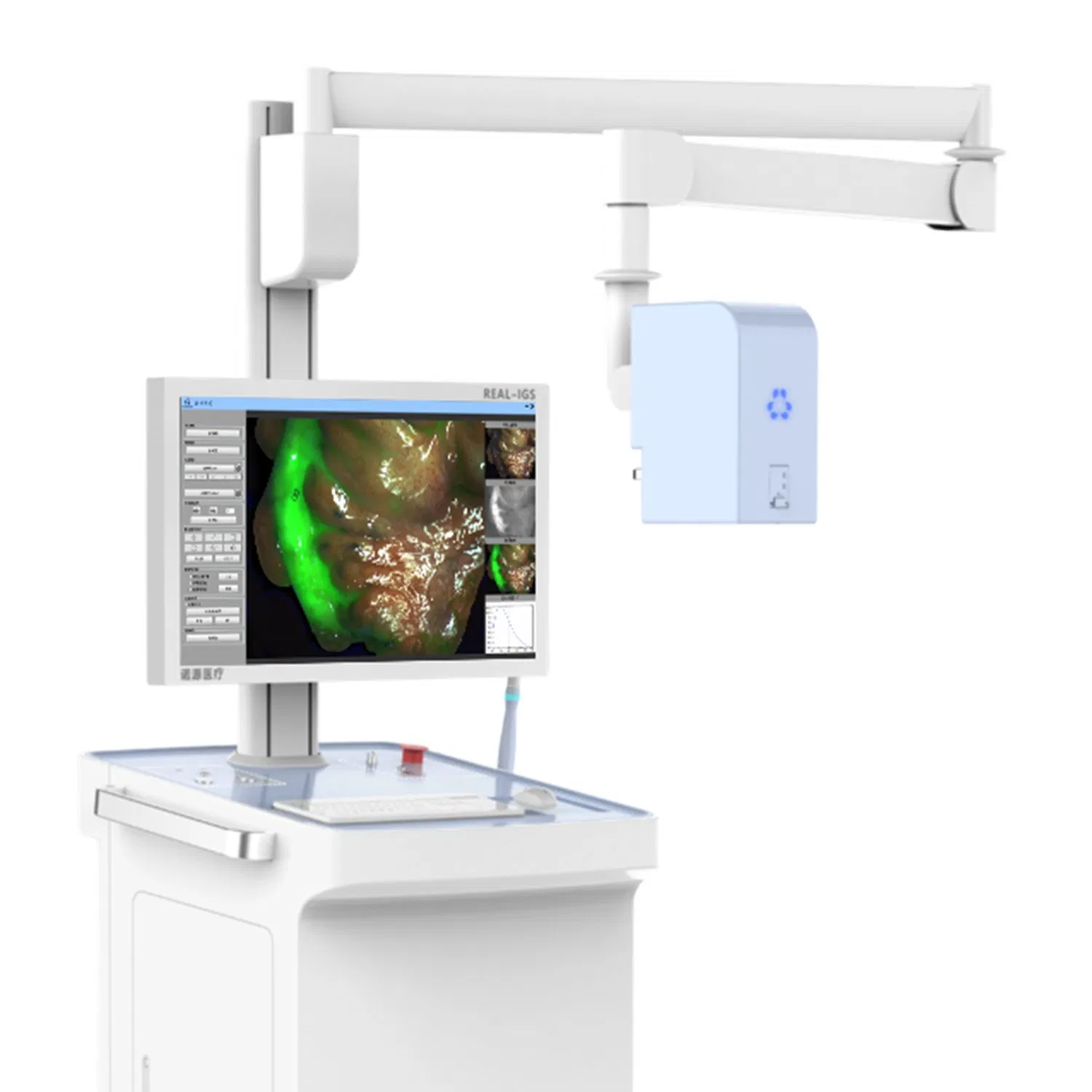
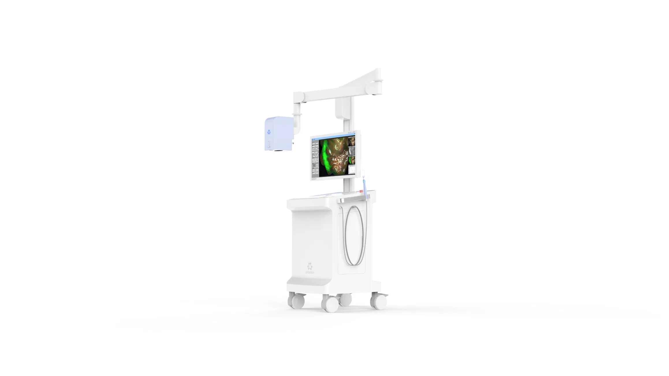
The FL1-10B surgical fluorescence imaging system is an advanced surgical guidance system employing a drug-device combination approach. Using Indocyanine Green (ICG) as a fluorescence probe, it leverages ultra-high sensitivity to identify submillimeter-sized tumors. It provides surgeons with high-definition visible light, fluorescence imaging, and quantitative diagnostic data in real-time.

This system is ideal for observing tumor tissues, margin assessments, blood supply (free skin flaps), lymph nodes, and complex anatomical structures like liver and lung segments.

⚙️

Technical Parameters

Project Content
Number of Camera Chips2CMOS
Convenient Handheld ProbeHandheld Spectral Quantitative Analysis Probe
Special Fluorescence DevelopmentAI Assisted Boundary Sharpening
Image Mode7 Types: White Light, Fluorescence, Fusion, Multimode, ColorGrading, Quantification, Spectroscopy
Laser Wavelength785nm
Fluorescence Detection Limit10-12M/L
Lens Zoom Factor4 Times
Focus ModeElectric Focus
Laser Grade3R
Software screenshot on the monitor
Surface light source
Handheld laser probe
🌟

Core Advantages

Product Benefits

  • Optimized for open surgery procedures.
  • Large field of view surface light source and unique handheld laser probe.
  • Ultra-high sensitivity (Surface source: nmol/L, Handheld probe: pmol/L).
  • Dual quantitative analysis system for objective data.

Technical Edge

  • Ultra-low Detection Limit: Real-time diagnosis of millimeter-sized tumors, enabling detection of residual cancer.
  • Quantitative Functions: ROI value and independent spectral digitization for precise clinical judgment.
  • Pseudo-color Grading: Visualizes the concentration gradient of tracers in target tissues.
🏥

Clinical Applications

Oral and Maxillofacial Surgery

Tumor localization
Tumor localization
Margin assessment
Margin assessment

Thyroid and Breast Surgery

Lymphatic mapping
Lymphatic mapping
Tumor identification

Dermatology and Plastic Surgery

Lower limb lymphatic mapping
Upper limb lymphatic mapping

General Surgery

Liver segment staining
Lymphatic tracing
Lymphatic tracing
Tumor identification
Margin determination

Frequently Asked Questions

1. What is the main use of the FL1-10B system? It is used for real-time surgical guidance to visualize blood flow, lymphatic systems, and tumor margins using ICG fluorescence.
2. What imaging modes does the system support? The system offers 7 modes: White Light, Fluorescence, Fusion, Multimode, Color Grading, Quantification, and Spectroscopy.
3. How sensitive is the fluorescence detection? It features ultra-high sensitivity, reaching down to 10-12 M/L, allowing for the identification of submillimeter-sized lesions.
4. Is the system suitable for thyroid surgery? Yes, it helps in identifying parathyroid glands and assessing their blood supply to reduce post-operative complications.
5. Does it provide quantitative analysis? Yes, it features a dual quantitative analysis system providing ROI values and independent spectral digitization for objective diagnosis.
6. Can it be used for flap transplantation? Absolutely. It is widely used to evaluate the blood supply and anastomosis of transplanted skin flaps in real-time.

Related Products